What is the human diaphragm and where is it located
What is the human diaphragm and where is it located
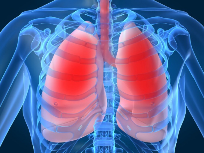
A diaphragm, also called "medullaryobstruction "is a muscular septum between the thoracic and abdominal cavity. It is a thin, wide, unpaired plate, curved convex side up and closing the lower opening of the chest. The diaphragm is only in mammals.

Where a person has a diaphragm
Conditionally, the diaphragm can be drawn along thethe lower edge of the ribs. The tip of the right dome of the diaphragm is usually at the level of the fourth intercostal space, the top of the left dome is at the level of the fifth. When breathing in, the diaphragm domes flatten and fall 2-3 cm. In the diaphragm, three parts are distinguished in the diaphragm: the sternum, the rib and the lumbar. From the posterior surface of the xiphoid process begins the sternal part. The widest rib part starts on the inner surface of the bone and cartilage parts of the six lower ribs. The lumbar part is divided into the right and left legs. Each of them comes from the anterolateral surface of 1-3 lumbar vertebrae and tendon lumbar-rib ligament.There are three holes in the diaphragm: aortic, esophageal and hollow vein.
Why you need a diaphragm
The diaphragm plays the role of the main respiratorymuscle. When contracted, it flattenes, which increases the volume of the chest and inspiration. When relaxed, the diaphragm assumes a convex spherical shape, which reduces the chest and provides exhalation.Involuntary trembling contractions of the diaphragm lead to hiccups.The diaphragm also performs a support (static)function, supporting the normal arrangement of the organs of the thoracic and abdominal cavities, contributes to the outflow of venous blood from the liver and the inferior vena cava to the heart and the movement of food through the esophagus.


