Tip 1: How does the procedure work?
Tip 1: How does the procedure work?
Magnetic resonance imaging is excellenta variant of diagnostic examination in various studies. This procedure is absolutely safe for patients, it can be repeated without fear. Thanks to MRI, you can put an accurate diagnosis and identify early changes in the body.

Instructions
1
Magnetic resonance therapy is not associated withionizing radiation, so the diagnosis does not harm the body. Despite absolute safety, this method has its own contraindications. Since the person with MRT has to be in a closed and narrow space, then people with claustrophobia procedure is contraindicated. Another limitation is the presence of metal objects (pacemakers, implants). Before recording on an MRI, the patient should notify the specialist about the presence of metal implants in the body. It is not recommended to conduct a similar examination during the first three months of pregnancy.
2
Magnetic resonance therapy can be performed onappointment of a doctor or on personal initiative. After the recording, the date of the MRI will be assigned, and you will be informed of the procedure. Before the therapy you will be given a special robe (you can stay in your clothes without the presence of metal zippers and buttons). Remove all accessories in the form of watches, jewelry, hairpins and hairpins, as well as a wig, hearing aid and denture.
3
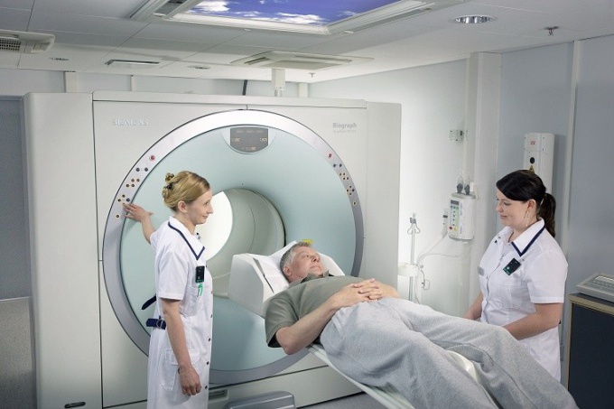
Metal objects can damageelectronics or disturb the magnetic field, which is used during the survey, the quality of the images may be poor. The tomograph contains a large tunnel-like magnet. The patient lays down on the moving table, which slowly moves into the tunnel. A magnetic field builds up elementary particles in the body. The collected signals are processed using a computer that produces a three-dimensional image of the body. Pictures are used for further examination, analysis and diagnosis.
4
Magnetic resonance therapy is absolutelypainless. You do not feel the radio waves and the magnetic field. Some pictures are obtained by introducing contrast through a vein in the hand. During the scan, strong signals are generated, repeated knocking and other noises, so you can enter into the ears cotton swabs. On average, the procedure lasts 2-15 minutes, some types of pictures are prepared within an hour. Breathing during the study should be calm, you can not move, the movements can distort the image of the pictures.
5
After the end of the survey you will be askedwait in the corridor until the pictures are completely ready and you will not be sure that you do not need additional results. Doctors will carefully analyze the images and write a research report, will put a conclusion. Sometimes they involve the discussion of the results of other doctors or the head of the department (in difficult cases).
Tip 2: MRI: preparation for the survey
The method of MRI - magnetic resonance therapy -based on the use of nuclear magnetic resonance, that is, the person does not receive the radiation load. Such a survey, according to medical professionals, is one of the safest kinds of diagnostics.

Take a sedative if you are afraid of a closedspace. Remove watches, jewelry, other metal objects, all this can affect the result of the survey. In addition, warn the doctor if there are implants in the body. Notify a specialist about the presence of tattoos - some types of paints include metal, and with MRI this will cause irritation on the skin.
When you are placed in a capsule of a tomograph,try to relax and lie still. Otherwise, the picture at the output will not be clear. The whole process is accompanied by loud noises, knocking and other noises. This is normal. With a doctor who conducts the procedure, you can talk through a microphone.
As soon as the scan is over, the doctor will examine the pictures and print out the conclusion. At your request, the results of MRI can be recorded on any external storage medium, for example, a disk or a USB flash drive.
Tip 3: How to get rid of myoma without surgery
Myoma - malignant formation in the cavityUterus, which is dangerous for the health and life of a woman, if not in time to not provide appropriate treatment. Modern medical technologies can cure the disease without surgical intervention. To do this, it is necessary to conduct a tumor investigation at a medical facility and follow the doctor's instructions clearly.

Diagnostics
Diagnosis of the disease is most often performed withusing ultrasound. Based on the findings, the doctor can prescribe a treatment, considering the patient's age, symptoms and ultrasound findings. On the basis of the available data, the possibility of hormonal therapy and the use of non-invasive methods of tumor treatment capable of preserving the organ, the health of the woman and even the reproductive function is determined.Ultrasound ablation
To remove myoma used ultrasoundablation, performed with a tomograph for MRI. The operation consists in the actual dissolution of the tumor with the help of ultrasonic waves. A complex of such operations is performed until the final result is achieved. The procedure is the gradual dissolution of small fragments of the tumor until all the affected cells are removed.The ablation method is remote and completely non-invasive.The effectiveness of this method is veryhigh. The possibility of use is due to the presence of contraindications for treatment, as well as the availability of the technical feasibility of this operation in each patient individually. The possibility of using ultrasound ablation can be determined by the location of the tumor in the uterus and the degree of development of the disease. However, ablation is often used in patients who have been shown to completely remove the uterus as the only way to get rid of the tumor. Against the background of this method of removal of myoma, medication is prescribed, which is also formed taking into account the patient's personal characteristics and will make the treatment more rapid. To determine the need for taking medications, a blood test can be assigned.
Embolization
Another new way to quickly get rid of fibroidsis embolization of arteries. Unlike ablation, this method is invasive, but no less effective. Its action is based on the isolation of the tumor from the vessels through which the malignant formation receives blood. After this, a special compound is inserted into the uterus, which clogs the blood vessels leading to the tumor. In the uterus, additional vessels are activated, through which the organ is supplied and after a few weeks the myoma gradually dies, and in its place connective tissue grows.This procedure is painless and is also one of the most effective methods of treating the disease.In Russia there are only a few clinics,capable of conducting similar operations to remove fibroids. Before treatment, you should consult about the possibility of the procedure in your hospital or decide on a trip to the nearest point where such high-tech methods are offered.


