How prenatal screening is done
How prenatal screening is done
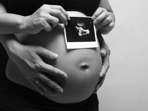
Prenatal screening is a complex of examinations that a woman undergoes between 11 and 13 weeks of pregnancy. Screening allows early detection of gross malformations of the fetus.

Every future mother wants her baby to behealthy, and pregnancy proceeded calmly and easily. Modern diagnostic techniques allow early pregnancy to identify serious pathologies and malformations of the fetus and make an idea of the health of the baby's future. In order to make sure that the baby is healthy and developing normally, all pregnant women should undergo prenatal screening. During the entire pregnancy period, screening is carried out three times.
First trimester screening
The most important is the very first screening, whichIs held at 11-13 weeks. In contrast to subsequent studies, it allows to recognize the signs of defects in the development of the nervous system, serious intrauterine malformations and genetic diseases, including Down syndrome, Edwards syndrome, Turner syndrome, and others. Prenatal screening of the first trimester consists of two stages - ultrasound examination of the fetus and biochemical screening. Biochemical research allows you to identify in the blood of a future mother specific substances that indirectly indicate the pathology of fetal development. If the woman is at risk in the analysis, she is assigned additional invasive examinations (chorion biopsy, amniocentesis, cordocentesis). The woman gives the biochemical blood test after the doctor has performed ultrasound. Ultrasound examination can reveal the most obvious violations in the development of the fetus, if they are present. Ultrasound also allows you to accurately calculate the timing of conception and make the necessary measurements for calculating risks. During the first screening, the doctor measures KTP (coccyx-parietal fetal size), BDP (biparietal size of head), and TVP (the thickness of the collar space). The increased thickness of the collar space indicates a high risk of the birth of a child with Down syndrome.Screening of the second and third trimesters
The second prenatal screening is conducted at 20-24Weeks of pregnancy in the middle of the second trimester. With his help, the doctor studies the anatomical development of the fetus and can in time recognize the malformations of the brain, heart, digestive tract, as well as the development of limbs and facial bones. Third prenatal screening is carried out in 30-32 weeks in the third trimester of pregnancy. It allows you to make sure of the normal course of pregnancy, to determine how the child is preparing for birth, and to identify some developmental defects that appear in later terms. If the defects found in the first and second trimester can be an indication for the artificial termination of pregnancy, the defects of the third trimester, as a rule, are corrected after the birth of the baby.


